Melatonin regulates the expression of Bone Morphogenetic Protein 15 (Bmp-15), Growth Differentiation Factor 9 (Gdf-9) and LH receptor (Lhr) genes in developing follicles of rats
Melatonin during the follicle development
Abstract
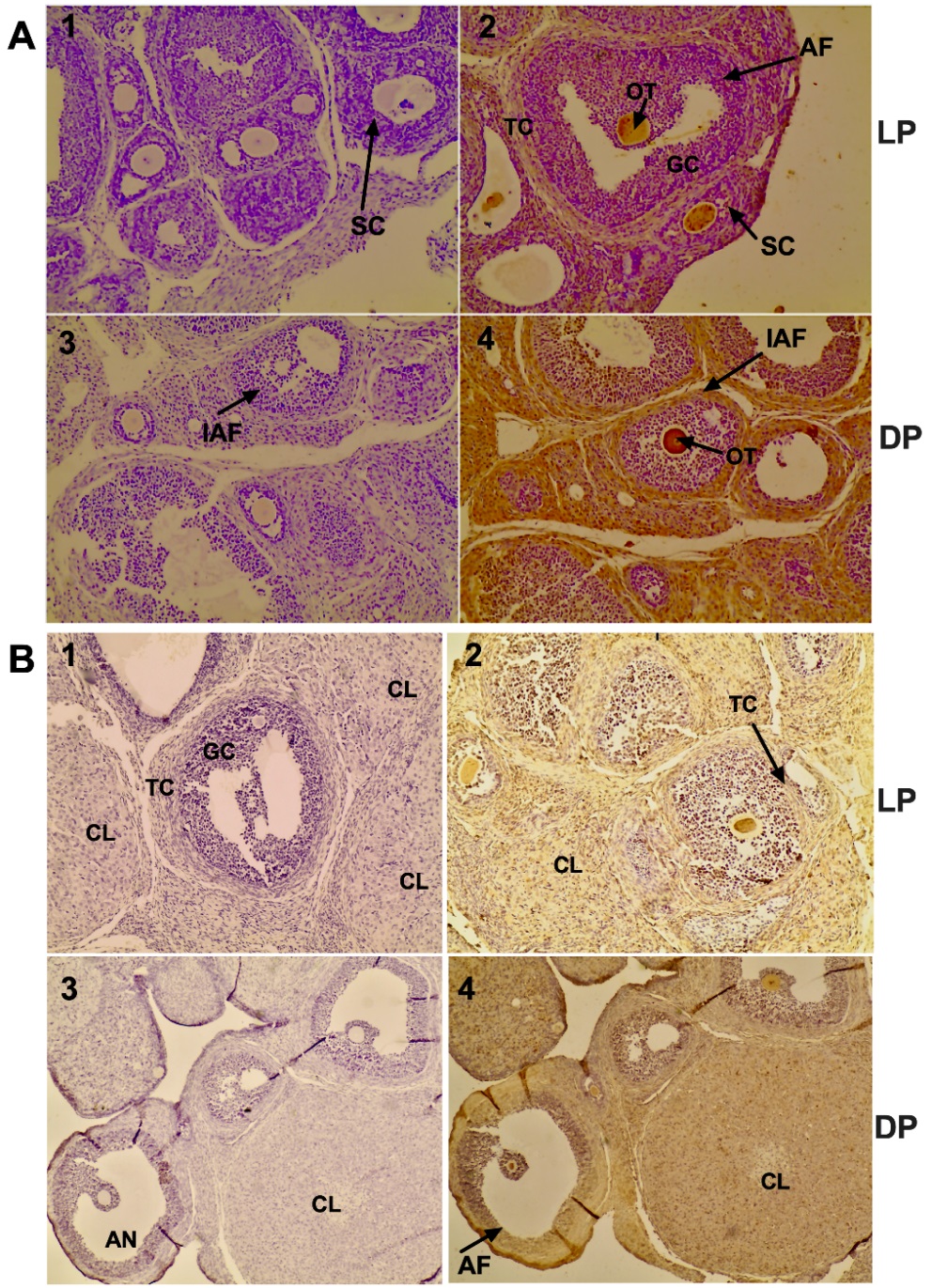
This study investigated the effects of melatonin supplementation on the daily mRNA expression of Bmp-15, Gdf-9, FSH (Fshr) and LH (Lhr) receptor genes and the effect of gonadotropin supplementation on protein expression of melatonin-synthetizing enzymes (ASMT and AANAT) during in vitro maturation (IMV) of rat follicle-enclosed oocytes. We also studied the effects of pinealectomy on the mRNA expression of Bmp-15, Gdf-9, Fshr and Lhr genes in immature oocytes and cumulus cells and on the protein expression and immunolocalization of AANAT and ASMT in immature ovaries. Ovaries were collected from sham-pinealectomized (INTACT) and pinealectomized (PINX) females during the light and dark phases of the 12h:12h light-dark cycle. Melatonin increased the Bmp-15 and Gdf-9 mRNA expression in follicle-enclosed oocytes during dark phase and Lhr mRNA expression during the light phase. The daily mRNA expression of Lhr was negatively correlated with the daily mRNA expression of Bmp-15 and Gdf-9 in follicle-enclosed oocytes supplemented with melatonin. There were no phase differences in oocytes mRNA expression of Bmp-15 and Gdf-9 from PINX females. Pinealectomy did not altered the mRNA expression of Lhr gene in cumulus cells. There were no phase differences in ASMT and AANAT expression in ovaries from INTACT and PINX females and their immunolocalization was seen in most of ovarian cells. AANAT expression in follicle-enclosed oocytes supplemented with LH from INTACT and PINX females was greater during the dark phase. The results suggest that melatonin regulates the daily Bmp-15, Gdf-9 and Lhr mRNA expression during the rat follicle development. There is also an indicative that melatonin plays an important role in the relationship between the daily expression of Lhr and the daily expression of Bmp-15 and Gdf-9 in follicle-enclosed oocytes during oocyte maturation process.
References
2. Nagyova E, Kalous J, Nemcova L (2016) Increased expression of pentraxin 3 after in vivo and in vitro stimulation with gonadotropins in porcine oocyte-cumulus complexes and granulosa cells. Dom. Anim. Endocrinol. 56: 29-35.
3. Epigg JJ. FSH (1979) stimulates hyaluronic acid synthesis by oocyte-cumulus cell complexes from mouse preovulatory follicles. Nature 281: 483-484.
4. Ge L, Sui H-S, Lan G-C, Liu N, Wang J-Z, Tan J-H (2008) Coculture with cumulus cells improves maturation of mouse oocytes denuded of the cumulus oophorus: observations of nuclear and cytoplasmic events. Fertil. Steril. 90: 2376-2388.
5. Ouandaogo ZG, Haouzi D, Assou S, Dechaud H, Kadoch IJ, Vos JD, Hamamah S (2011) Human cumulus cells molecular signature in relation to oocyte nuclear maturity stage. Plos One 6: e27179.
6. Robert C, Gagné D, Lussier JG, Bousquet D, Barnes FL, Sirard M-A (2003) Presence of LH receptor mRNA in granulosa cells as a potential marker of oocyte developmental competence and characterization of the bovine splicing isoforms. Reproduction 125: 437-446.
7. Su Y-Q, Wu X, O’Brien MJ, Pendola FL, Denegre JN, Matzuk MM, Eppig JJ (2004) Synergistic roles of BMP15 and GDF9 in the development and function of the oocyte-cumulus cell complex in mice: genetic evidence for an oocyte-granulosa cell regulatory loop. Dev. Biol. 276: 64-73.
8. Otsuka F, McTavish K, Shimasaki S (2011) Integral role of GDF-9 and BMP-15 in ovarian function. Mol. Reprod. Dev. 78: 9-21.
9. Castro FC, Cruz MHL, Leal CLV (2016) Role of growth differentiation factor 9 and Bone morphogenetic protein 15 in ovarian function and their omportance in mammalian female fertility – A review. Asian Australas. J. Anim. Sci. 29: 1065-1074.
10. Elvin JA, Clark AT, Wang P, Wolfman NM, Matzuk MM (1999) Paracrine actions of growth differentiation factor-9 in the mammalian ovary. Mol. Endocrinol. 13: 1033-1048.
11. Garcia P, Aspee K, Ramirez G, Dettleff P, Palomino J, Peralta OA, Parraguez VH, De los Reyes M (2019) Influence of growth differentiation factor-9 and bone morphogenetic protein 15 on in vitro maturation of canine oocytes. Reprod. Dom. Anim. 54: 373-380.
12. Velásquez A, Mellisho E, Castro FO, Rodríguez-Álvarez L (2019) Effect of BMP15 and or AMH during in vitro maturation of oocytes from involuntarily culled dairy cows. Reprod. Dom. Anim. 84: 209-223.
13. Moore RK, Shimazaki S (2005) Molecular biology and physiological role of the oocyte factor, BMP-15. Mol. Cell. Endocrinol. 234: 67-73.
14. Vitt UA, Hayashi M, Klein C, Hsueh AJW (2000) Growth differentiation factor-9 stimulates proliferation but suppresses the follicle-stimulating hormone-induced differentiation of cultured granulosa cells from small antral and preovulatory rat follicles. Biol. Reprod. 62: 370-377.
15. Cipolla-Neto J, Amaral FG (2018) Melatonin as a hormone: new physiological and clinical insights. Endocr. Rev. 39: 990-1028.
16. Tamura H, Nakamura Y, Korkmaz A, Manchester LC, Tan D-X, Sugino N, Reiter RJ (2009) Melatonin and the ovary: physiological and pathophysiological implications. Fertil. Steril. 92: 328-343.
17. Tamura H, Takasaki A, Taketani T, Tanabe M, Kizuka F, Lee L, Tamura I, Maekawa R, Asada H, Yamagata Y, Sugino N (2012) The role of melatonin as an antioxidant in the follicle. J. Ovarian Res. 5: 5. doi:10.1186/1757-2215-5-5.
18. Maganhin CC, Fuchs LFP, Simões RS, Oliveira-Filho RM, Simões MJ, Baracat EC, Soares JM Jr (2013) Effects of melatonin on ovarian follicles. Eur. J. Obstet. Gynecol. Reprod. Biol. 166: 178-184.
19. Tamura H, Takasaki A, Miwa I, Taniguchi K, Maekawa R, Asada H, Taketani T, Matsuoka A, Yamagata Y, Shimamura K, Morioka H, Ishikawa H, Reiter RJ, Sugino N (2008) Oxidative stress impairs oocyte quality and melatonin protects oocytes from free radical damage and improves fertilization rate. J. Pineal Res. 44: 280-287.
20. Wang Y, Zeng S (2018) Melatonin promotes ubiquitination of phosphorylated pro-apoptotic protein Bcl-2-interacting mediator of cell death-extra long (BimEL) in porcine granulosa cells. Int. J. Mol. Sci. 19: 3431. doi:10.3390/ijms19113431.
21. Sakaguchi K, Itoh MT, Takahashi N, Tamuri W, Ishizuka B (2013) The rat oocyte synthesizes melatonin. Reprod. Fertil. Dev. 25: 674-682.
22. He C, Wang J, Zhang Z, Yang M, Li Y, Tian X, Ma T, Tao J, Zhu K, Song Y, Ji P, Liu G (2016) Mitochondria synthesize melatonin to ameliorate its function and improve mice oocyte’s quality under in vitro conditions. Int. J. Mol. Sci. 17: 939. doi:10.3390/ijms17060939.
23. Itoh MT, Ishizuka B, Kuribayashi Y, Amemiya A, Sumi Y (1999) Melatonin, its precursors, and synthesizing enzyme activities in the human ovary. Mol. Hum. Reprod. 5: 402-408.
24. An Q, Peng W, Cheng Y, Lu Z, Zhou C, Zhang C, Su J (2019) Melatonin supplementation during maturation of oocyte enhances subsequent development of bovine cloned embryos. J. Cell Physiol. 234: 17370-17381.
25. Woo MMM, Tai C-J, Kang SK, Nathwani PS, Pang SF, Leung PCK (2011) Direct action of melatonin in human granulosa-luteal cells. J. Clin. Endocrinol. Metab. 2086: 4789-4797.
26. Nakamura E, Otsuka F, Terasaka T, Inagaki K, Hosoya T, Tsukamoto-Yamauchi N, Toma K, Makino H (2014) Melatonin counteracts BMP-6 regulation of steroidogenesis by rat granulosa cells. J. Steroid Biochem. Mol. Biol. 143: 233-239.
27. Otsuka F (2018) Interaction of melatonin and BMP-6 in ovarian steroidogenesis. Vitam. Horm. 107: 137-153.
28. Simonneaux V, Ribelayga C (2003) Generation of the melatonin endocrine message in mammals: a review of the complex regulation of melatonin synthesis by norepinephrine, peptides, and other pineal transmitters. Pharmacol. Rev. 55: 325-395.
29. Hillensjö T, LeMaire WJ (1980) Gonadotropin releasing hormone agonists stimulate meiotic maturation follicle-enclosed rat oocytes in vitro. Nature 287: 145-146.
30. Sela-Abramovich S, Chorev E, Galiani D, Dekel N (2005) Mitogen-activated protein kinase mediates luteinizing hormone-induced breakdown of communication and oocyte maturation in rat ovarian follicles. Endocrinology 146: 1236-1244.
31. Livak KJ, Schmittgen TD (2001) Analysis of relative gene expression data using real-time quantitative PCR and the 2-ΔΔCT method. Methods 25: 402-408.
32. Vandesompele J, De Preter K, Pattyn F, Poppe B, Roy NV, Paepe AD, Speleman F (2002) Accurate normatization of real-time quantitative RT-PCR data by geometric averaging of multiple internal control genes. Genome Biol. 3: RESEARCH0034.
33. Amaral FG, Turati AO, Barone M, Scialfa JH, Buonfiglio DC, Peres R, Peliciari-Garcia RA, Afeche SC, Lima L, Scavone C, Bordin S, Reiter RJ, Cipolla-Neto J. (2014) Melatonin synthesis impairment as new deleterious outcome of diabetes-derived hyperglycemia. J. Pineal Res. 57: 67-79.
34. Hoffman RA, Reiter RJ (1965) Rapid pinealectomy in hamsters and other small rodents. Anat. Rec. 153: 19-21.
35. Coelho LA, Andrade-Silva J, Motta-Teixeira LC, Amaral FG, Reiter RJ, Cipolla-Neto J (2019) The absence of pineal melatonin abolishes the daily rhythm of Tph1 (Tryptophan Hydroxylase 1), Asmt (Acetylserotonin O-Methyltransferase) and Aanat (Aralkylamine N- Acetyltransferase) mRNA expressions in rat testes. Mol. Neurobiol. 56: 7800-7809.
36. Wei L-N, Huang R, Li L-L, Fang C, Li Y, Liang X-Y (2014) Reduced and delayed expression of GDF9 and BMP15 in ovarian tissues from women with polycystic ovary syndrome. J. Assit. Reprod. Genet. 31: 1483-1490.
37. Wei L-N, Fang C, Huang R, Li L, Zhang M, Liang X (2012) Change and significance of growth differentiation factor 9 and bone morphogenetic protein15 expression during oocyte maturation in polycystic ovary syndrome with ovarian stimulation. Zhonghua Fu Chan Ke Za Zhi. 47 (11): 818-22.https://pubmed.ncbi.nlm.nih.gov/23302121.
38. Teixeira Filho FL, Baracat EC, Lee TH, Suh CS, Matsui M, Chang RJ, Shimasaki S, Erickson GF (2002) Aberrant expression of growth differentiation factor-9 in oocytes of women with polycystic ovary syndrome. J. Clin. Endocrinol. Metab. 87: 1337-1344.
39. Zhao S-Y, Qiao J, Chen Y-J, Liu P, Li J, Yan J (2010) Expression of growth differentiation factor-9 and bone morphogenetic protein-15 in oocytes and cumulus granulosa cells of patients with polycystic ovary syndrome. Fertil. Steril. 94: 261-267.
40. Beli M, Shimasaki S (2018) Molecular Aspects and clinical relevance of GDF9 and BMP15 in ovarian function. Vitam. Horm. 107: 317-348.
41. Qiao J, Feng HL (2011) Extra- and intra-ovarian factors in polycystic ovary syndrome: impact on oocyte maturation and embryo developmental competence. Hum. Reprod. Update 17: 17-33.
42. Kollmann M, Martins WP, Lima MLS, Craciunas L, Nastri CO, Richardson A, Raine-Fenning N (2016) Strategies for improving outcome of assisted reproduction in women with polycystic ovary syndrome: Systematic review and meta-analysis. Ultrasound Obstet. Gynecol. 48: 709-718.
43. Nikmard F, Hosseini E, Bakhtiyari M, Ashrafi M, Amidi F, Aflatoonian R (2017) Effects of melatonin oocyte maturation in PCOS mouse model. Anim. Sci. J. 88: 586-592.
44. Kim MK, Park EA, Kim HJ, Choi WY, Cho JH, Lee WS, Cha KY, Kim YS, Lee DR, Yoon TK (2013) Does supplementation of in-vitro culture medium with melatonin improve IVF outcome in PCOS? Reprod. Biomed. Online 26: 22-29.
45. Riaz H, Yousuf MR, Liang A, Hua GH, Yang L (2019) Effect of melatonin on regulation of apoptosis and steroidogenesis in culture buffalo granulosa cells. Anim. Sci. J. 90: 473-480.
46. Nagyova E, Kalous J, Nemcova L (2016) Increased expression of pentraxin 3 after in vivo and in vitro stimulation with gonadotropins in porcine oocyte-cumulus complexes and granulosa cells. Dom. Anim. Endocrinol. 56: 29-35.
47. Epigg JJ (1979) FSH stimulates hyaluronic acid synthesis by oocyte-cumulus cell complexes from mouse preovulatory follicles. Nature 281: 483-484.
48. Nakamura K, Minegishi T, Takakura Y, Miyamoto K, Hasegawa Y, Ibuki Y, Igarashi M (1991) Hormonal regulation of gonadotropin receptor mRNA in rat ovary during follicular growth and luteinization. Mol. Cell. Endocrinol. 82: 259-263.
49. Walker RF, McCamant S, Timiras PS (1982) Melatonin and the influence of the pineal gland on timing of the LH surge in rats. Neuroendocrinology 35: 37-42.
50. Chiba A, Akema T, Toyoda J (1994) Effects of pinealectomy and melatonin on the timing of proestrous luteinizing hormone surge in the rat. Neuroendocrinology 59: 163-168.
51. Acuña-Castroviejo D, Escames G, Venegas C, Díaz-Casado ME, Lima-Cabello E, López LC, Rosales-Corral S, Tan D-X, Reiter RJ (2014) Extrapineal melatonin: sources, regulation, and potential functions. Cell. Mol. Life Sci. 71: 2997-3025.
52. Stefulj J, Hörtner M, Ghosh M, Schauenstein K, Rinner I, Wölfler A, Semmier J, Liebmann M (2001) Gene expression of the key enzymes of melatonin synthesis in extrapineal tissues of the rat. J. Pineal Res. 30: 243–247.
53. Reiter RJ, Rosales-Corral S, Tan DX, Jou MJ, Galano A, Xu B (2017) Melatonin as a mitochondria-targeted antioxidant: one of evolution’s best ideas. Cell. Mol. Life Sci. 74: 3863-3881.
54. Dardes RC, Baracat EC, Simões MJ (2000) Modulation of estrous cycle and LH, FSH and melatonin levels by pinealectomy and sham-pinealectomy female rats. Prog. Neuro-Psychopharmacol. Biol. Psychiat. 24: 441-453.
55. Chuffa LGA, Seiva FRF, Fávaro WJ, Teixeira GR, Amorim JPA, Mendes LO, Fioruci BA, Pinheiro PFF, Fernandes AAH, Franci JAA, Dellela FK, Martinez M, Martinez FE (2011) Melatonin reduces LH, 17 beta-estradiol and induces differential regulation of sex steroid receptors in reproductive tissues during rat ovulation. Reprod. Biol. Endocrinol. 9: 108.
56. Soares JM, Simões MJ, Oshima CTF, Mora OA, Lima GR, Baracat EC (2003) Pinealectomy changes rat ovarian interstitial cell morphology and decreases progesterone receptor expression. Gynecol. Endocrinol. 17: 115-123.
57. Barbacka-Surowiak G, Surowiak J, Stoklosowa S (2003) The involvement of the suprachiasmatic nuclei in the regulation of estrous cycles in rodents. Reprod. Biol. 3: 99-129.
58. Coelho LA, Peres R, Amaral FG, Reiter RJ, Cipolla-Neto J (2015) Daily differential expression of melatonin-related genes and clock genes in rat cumulus-oocyte complex: Changes after pinealectomy. J. Pineal Res. 58: 490-499.
59. He C, Ma T, Shi J, Zhang Z, Wang J, Zhu K, Li Y, Yang M, Song Y, Liu G (2016) Melatonin and its receptor MT1 are involved in the downstream reaction to luteinizing hormone and participate in the regulation of luteinization in the different species. J. Pineal Res. 61: 279-290.
60. Teixeira AAC, Simões MJ, Teixeira VW, Soares JM Jr (2004) Evaluation of the implantation in pinealectomized and/or submitted to the constant illumination rats. Int. J. Morphol. 22: 189-194.
61. Dair EL, Simões RS, Simões MJ, Romeu LRG, Oliveira-Filho RM, Haidar MA, Baracat EC, Soares JM Jr (2008) Effects of melatonin on the endometrial morphology and embryo implantation in rats. Fertil. Steril. 89: 1299-1305.
62. Manganhin CC, Simões RS, Fuchs LFP, Sasso GRS, Simões MJ, Baracat EC, Soares JM Jr (2014) Melatonin influences on steroidogenic gene expression in the ovary of pinealectomized rats. Fertil. Steril. 102: 291-297.
63. Tamura H, Jozaki M, Tanabe M, Shirafuta Y, Mihara Y, Shinagawa M, Tamura I, Maekawa R, sato S, Taketani T, Takasaki A, Reiter RJ, Sugino N (2020) Importance of melatonin in assisted reproductive technology and ovarian age. Int. J. Mol. Sci. 21: 1135. doi:10.3390/ijms21031135.


This work is licensed under a Creative Commons Attribution 4.0 International License.
For all articles published in Melatonin Res., copyright is retained by the authors. Articles are licensed under an open access Creative Commons CC BY 4.0 license, meaning that anyone may download and read the paper for free. In addition, the article may be reused and quoted provided that the original published version is cited. These conditions allow for maximum use and exposure of the work, while ensuring that the authors receive proper credit.
In exceptional circumstances articles may be licensed differently. If you have specific condition (such as one linked to funding) that does not allow this license, please mention this to the editorial office of the journal at submission. Exceptions will be granted at the discretion of the publisher.


